What is cancer?
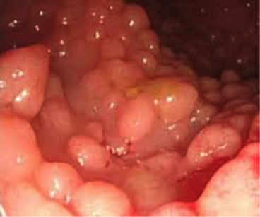
Picture of Colon Cancer: Found at https://familyhistorybowelcancer.wordpress.com/tag/attenuated-familial-adenomatous-polyposis/
|
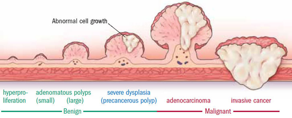
Cancer is the abnormal growth of cells within the body and they are many factors, genetic or environmental that causes them to become malignant. While cancer is stealing lives every year, we have still not been able to come up with a way to effectively treat this disease, besides chemotherapy, radiation and surgery [1].
Whether toxins, chemicals, exposure to sunlight, radiation or genetic predispositions cause cancer, every way and every cancer starts with cell division failures and overgrowth [2]. This creates cells that are abnormal in size, genetic structure and an overexpression of many proteins. This can be seen in both of the videos below [3,4]. |
What is colorectal cancer?
Colorectal Cancer is the 3rd deadliest of all cancers, only preceded by lung and bronchial cancer. It causes around 50,000 death every year, even though it is very preventable. It occurs when malignant cells form within the keratinocytes or the intestinal epidermal cells that line the colon as pictured below [6].
Cancers in the colon generally start as a polyp, which can be benign or eventually become malignant and growth unregulated into large masses, as seen below [7]. Since it is not a cancer that can be seen with the naked eye, there are many symptoms of colon cancer such as abdominal pain and blood in the stool that are warning signs, in which a colonoscopy should occur. These symptoms are explained in more detail in the video to the left [8].
What is a keratinocyte?
|
Keratinocytes are the most common cell type of the epidermal layer and lines all major organs in the body. These cells also aid in cancer transport because they have large direct outflows to the bloodstream which enables spread. Keratinocytes in the intestines are called intestinal epidermal cells, which are pictured below. Keratinocytes main function is a barrier to outside invasions and are important for hair and nail based development. This is why when phenotypes of various model organisms are explored, we see that they develop deficiencies in their keratinocytes, with undeveloped hair patches, brittle skin and cracked nails.
|
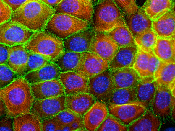
Picture of Keratinocytes. Found at http://cellntec.com/products/skin/keratinocyte-cells/
|
What Is 14-3-3sigma?
While many genes could potentially be mutated to lead to cancer, one particular family of seven proteins called 14-3-3 proteins have the highest affinity for cancers. They are incredibly important in cellular function and homeostasis as well in that the loss of one of the 7 14-3-3's is generally tolerable, but a loss of more than one will result in death for eukaryotes. Each 14-3-3 protein has different in function and the others are gamma, beta, epsilon, zeta, eta and tau, but all are involved in cell cycle regulation, apoptosis and translation [9]. One of the proteins, 14-3-3sigma is known to play an important role in many different cancers but I chose to research a disease that has personal significance to me, which you can read about here.
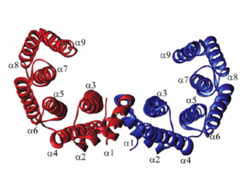
The 14-3-3 proteins are a large family of approximately 30kDa acidic proteins which exist primarily as homo- and heterodimeric within all eukaryotic cells [(PUBMED:1671102), (PUBMED:11911880)]. The different 14-3-3 proteins are all involved in the cell cycle, translation and apoptosis [9]. Each 14-3-3 protein can has the same basic structure, as described in this image, a different N and C terminus and a well conserved middle, which is congruent to the 14-3-3 protein domain that is well conserved across many organisms. The picture to the right explains this structure [12].
The monomer consists of nine helices organised in an antiparallel manner, forming an L-shaped structure. The interior of the L-structure is composed of four helices: H3 and H5, which contain many charged and polar amino acids, and H7 and H9, which contain hydrophobic amino acids. These four helices form the concave amphipathic groove that interacts with target peptides [13].
14-3-3 proteins mainly bind proteins containing phosphothreonine or phosphoserine motifs however exceptions to this rule do exist. Proteins appear to initially bind to a single dominant site and then subsequently to many, much weaker secondary interaction sites. The 14-3-3 dimer is capable of changing the conformation of its bound ligand whilst itself undergoing minimal structural alteration [13].
Where is 14-3-3sigma located in the cell?
The 14-3-3sigma gene is located on 1st Chromosome at the 1p36.11 location, as you can see in the image below [10]. It is 1,336 bp long and has only one exon (Accession: NM_006142.3.). This encodes for 248 amino acids, (Accession Number: CAG46703.1) creating the 14-3-3sigma protein [11].
Its found in various cell types as well and localizes within the nucleus of the cell.
Its found in various cell types as well and localizes within the nucleus of the cell.
What is the Function of 14-3-3Sigma?
14-3-3sigma has the most recognized effects in cancer of all types, so we know it plays a huge role in the pathways leading to this disease [11].
Mutations in this protein that lead to cancer are caused by hyper-methylation of the promoter region of the protein, which creates a loss of function discrepancy. The loss of the 14-3-3sigma protein results in tumors, so research has stated that 14-3-3sigma has a tumor-suppression function within the cell, and it is also known to "establish a connection between the aberrant regulation of mitotic translation and improper cytokinesis resulting in a possible cancer [11]."
While much is known about this protein in general, its specific role in cell division is still relatively unknown.
Videos
|
|
|
|
|
|
References:
[1] Colon and Rectal Cancer. (n.d.). Retrieved February 19, 2015, from http://www.cancer.gov/cancertopics/types/colon-and-rectal
[2] Board, A. (n.d.). Retrieved February 19, 2015, from http://www.ncbi.nlm.nih.gov/pubmedhealth/PMH0002267/
[3] Youtube Video: Cancer Unregulated Cell Growth. https://www.youtube.com/watch?v=IeUANxFVXKc
[4] Youtube Video: HeLa Cell Divison.
https://www.youtube.com/watch?v=XSOjy906MuQ
[5] Chan, A. (2010, September 10). The 10 Deadliest Cancers and Why There's No Cure. Retrieved February 19, 2015, from http://www.livescience.com/11041-10-deadliest-cancers-cure.html
[6] Picture of Keratinocytes: http://iwebpd.saschina.org/stwebpd/iris01pd2015/Digestive_System/Small_Intestine.html
[7] Pictures of Cancer Polyps: http://www.askdoctork.com/do-i-have-colon-cancer-if-my-doctor-found-a-polyp-during-my-colonoscopy-201402156075
[8] Youtube Video: Symptoms of Colon Cancer: https://www.youtube.com/watch?v=LfXHoSVXetQ
[9] Hermeking, H. (2003). The 14-3-3 cancer connection. Nature Reviews Cancer, 3(12), 931-943. doi: 10.1038/nrc1230.
[10] Picture of Chromosome 1: http://commons.wikimedia.org/wiki/File:Chromosome1.PNG
[11] Wilker, E. W., van Vugt, M., Artim, S. A., Huang, P. H., Petersen, C. P., Reinhardt, H. C., . . . Yaffe, M. B. (2007). 14-3-3 sigma controls mitotic translation to facilitate cytokinesis. Nature, 446(7133), 329-332. doi: 10.1038/nature05584
[12] 14-3-3sigma structure. Retrieved on: http://www.mpg.de/826613/forschungsSchwerpunkt
[13] 14-3-3sigma function. Retrieved on: http://www.uniprot.org/uniprot/P31947#interaction
